The current examine investigated the impact of tetrandrine on lung most cancers cell progress and apoptosis, and its attainable underlying molecular mechanism.
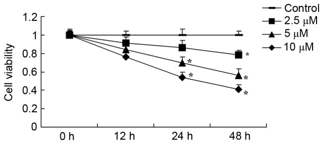
A549 human lung most cancers cells have been incubated with between 2.5 and 1zero µM tetrandrine for 12, 24 and 48 h, following which the impact of tetrandrine on cell viability and apoptosis have been assessed utilizing an MTT assay and circulation cytometry.
ELISA and western blotting have been used to research VEGF exercise, and the expression of poly (ADP-ribose) polymerase (PARP), phosphorylated protein kinase B (Akt), Bcl-2-associated X protein (Bax), hypoxia inducible issue (HIF)-1α and inter–mobile adhesion molecule–1 (ICAM-1). Tetrandrine successfully suppressed the expansion of and induced apoptosis in A549 lung most cancers cells.
The expression of PARP, Bax, intercellular adhesion molecule–1 (ICAM-1) and vascular endothelial progress issue (VEGF) was considerably upregulated, and the phosphorylation of Akt and expression of HIF-1α was considerably suppressed in A549 lung most cancers cells.
Subsequently, tetrandrine could suppress cell viability and induce apoptosis through the VEGF/HIF-1α/ICAM-1 signaling pathway.
Inducible indoleamine 2,3-dioxygenase 1 and programmed dying ligand 1 expression because the efficiency marker for mesenchymal stromal cells.
OBJECTIVEEstablishment of a efficiency assay within the manufacturing of clinical-grade mesenchymal stromal cells (MSCs) has been a problem on account of problems with relevance to perform, timeline and variability of responder cells.
On this examine, we tried to develop a efficiency assay for MSCs.METHODSClinical-grade bone marrow-derived MSCs have been manufactured. The phenotype and immunosuppressive capabilities of the MSCs have been evaluated based mostly on the Worldwide Society for Mobile Remedy pointers.
Resting MSCs licensed by interferon (IFN)-γ publicity in a single day have been evaluated for modifications in immune suppression and immune-relevant proteins.
The connection of immune-relevant protein expression with immunosuppression of MSCs was analyzed.RESULTSMSC supressed third-party T-lymphocyte proliferation with excessive inter-donor and inter-test variability.
The suppression of T-lymphocyte proliferation by IFN-γ-licensed MSCs correlated with that by resting MSCs. Many mobile proteins have been up-regulated after IFN-γ publicity, together with indoleamine 2,3-dioxygenase 1 (IDO-1), programmed dying ligand 1 (PD-L1), vascular cell adhesion molecule 1 (VCAM-1), intercellular adhesion molecule 1 (ICAM-1) and bone marrow stromal antigen 2 (BST-2).
The expression ranges of IDO-1 and PD-L1 on licensed MSCs, not VCAM-1, ICAM-1 or BST-2 on licensed MSCs, correlated with MSC suppression of third-party T-cell proliferation.
CONCLUSIONSA circulation cytometry-based assay of MSCs post-IFN-γ publicity measuring expression of intracellular protein IDO-1 and cell floor protein PD-L1 captures two mechanisms of suppression and provides the potential of a related, speedy assay for MSC-mediated immune suppression that will match with the manufacturing course of.